The maxillofacial region is a dynamic anatomical zone where dental, skeletal, and systemic interactions converge. While odontogenic cysts are among the most commonly discussed jaw lesions, a broader range of bone pathologies exists that demands deeper attention. Conditions such as fibro-osseous lesions, giant cell granulomas, and benign and malignant bone tumors represent a diverse and diagnostically challenging subset of maxillofacial abnormalities. These lesions vary widely in etiology, clinical behavior, radiographic appearance, and histological architecture. Their accurate identification and management require a multidisciplinary approach informed by clinical, radiologic, and pathologic correlation. This comprehensive understanding is especially relevant to specialists like Eric Starley DMD, whose focus on precision in diagnosis and surgical treatment underlines the importance of nuanced interpretation in jaw pathology.
Fibro-Osseous Lesions: A Diagnostic Conundrum
Fibro-osseous lesions of the jaws are a group of conditions characterized by the replacement of normal bone with fibrous tissue containing varying amounts of mineralized material. These include fibrous dysplasia, ossifying fibroma, and cemento-osseous dysplasia. While they share some histologic features, their clinical presentation and behavior differ significantly.
Fibrous dysplasia typically affects younger patients and can involve one (monostotic) or multiple (polyostotic) bones. In the jaws, it presents as a painless swelling with a classic “ground-glass” radiographic appearance. Histologically, it features irregular trabeculae of woven bone within a fibrous stroma, often described as “Chinese characters.”
Ossifying fibroma, on the other hand, is a true neoplasm. It appears well-circumscribed radiographically and demonstrates a centrifugal growth pattern. Its internal structure varies from radiolucent to radiopaque depending on the degree of mineralization. Microscopically, it shows a more cellular stroma with ossicles of woven and lamellar bone.
Cemento-osseous dysplasia, commonly seen in middle-aged African American women, often arises in the periapical regions of the anterior mandible. It is usually asymptomatic and discovered incidentally. Its progression from radiolucent to mixed and eventually radiopaque lesions helps distinguish it radiographically.
Differentiating among these entities is critical, as treatment approaches range from conservative monitoring to surgical excision. Misdiagnosis can lead to unnecessary interventions or neglect of progressive disease.
Central Giant Cell Granulomas: Aggressive Yet Benign
Central giant cell granulomas (CGCG) are benign but potentially aggressive lesions that typically occur in the anterior mandible of individuals under 30 years of age. They may present as painless swellings or be associated with pain and cortical expansion. Radiographically, CGCGs appear as unilocular or multilocular radiolucencies with well-defined but often scalloped borders. Root resorption and displacement of teeth are common features.
Histologically, CGCGs consist of multinucleated giant cells dispersed within a background of spindle-shaped mesenchymal cells and hemorrhagic stroma. The number and distribution of giant cells can vary, and areas of reactive bone formation may be present.
The biological behavior of CGCGs ranges from indolent to aggressive. Aggressive lesions demonstrate rapid growth, root resorption, cortical perforation, and recurrence after curettage. As such, treatment strategies may include intralesional corticosteroid injections, calcitonin therapy, or surgical resection depending on lesion size and aggressiveness.
Differential diagnosis includes brown tumors of hyperparathyroidism, cherubism, and aneurysmal bone cysts. Biochemical testing for calcium and parathyroid hormone levels is essential when hyperparathyroidism is suspected. Accurate diagnosis hinges on a combination of clinical, radiologic, histologic, and laboratory findings.
Benign Bone Tumors: Discrete Yet Disruptive
Benign bone tumors such as osteomas, osteoblastomas, and cementoblastomas present unique challenges in diagnosis and management. Although non-malignant, their location and growth patterns can have significant functional and esthetic implications.
Osteomas are slow-growing lesions composed of compact or cancellous bone. They are commonly found on the surfaces of craniofacial bones and are often associated with Gardner syndrome when multiple osteomas are present. Radiographically, they appear as dense, well-circumscribed radiopaque masses.
Osteoblastomas are rare lesions typically occurring in the posterior mandible. They present as painful swellings and may appear radiolucent or mixed on imaging. Histologically, they consist of irregular trabeculae of osteoid and woven bone with prominent osteoblastic rimming.
Cementoblastomas arise from the cementum-producing cells and are often attached to the roots of mandibular molars. They manifest radiographically as well-defined radiopaque masses with a radiolucent halo. Unlike other benign lesions, cementoblastomas often cause pain and require surgical extraction of the involved tooth.
These tumors, although rare, can mimic other jaw lesions both clinically and radiographically. Thorough histopathological analysis is essential to confirm diagnosis and plan appropriate management.
Malignant Bone Tumors: Uncommon But Critical
Malignant tumors of the jawbone, such as osteosarcomas and chondrosarcomas, are rare but serious conditions requiring immediate attention. Osteosarcoma is the most common primary malignant bone tumor of the jaws and typically presents in the third to fourth decade of life, earlier than its long bone counterpart.
Clinically, osteosarcomas manifest as painful swellings with rapid growth, tooth mobility, and paresthesia. Radiographic findings vary but may include mixed radiolucent-radiopaque lesions with poorly defined borders and a characteristic “sunburst” periosteal reaction.
Histologically, osteosarcomas demonstrate malignant mesenchymal cells producing osteoid matrix. The degree of differentiation and subtype (osteoblastic, chondroblastic, fibroblastic) influence prognosis and treatment planning.
Chondrosarcomas, arising from cartilaginous tissue, are less common but often affect the anterior maxilla. They are slow-growing but locally destructive. Imaging reveals lobulated radiolucent lesions with areas of calcification, and histology confirms the presence of malignant chondrocytes within a cartilaginous matrix.
Management of malignant bone tumors typically involves surgical resection with wide margins, often followed by adjunctive chemotherapy or radiotherapy. Early detection and multidisciplinary management are essential for improving survival rates.
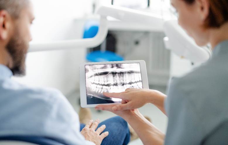
Imaging Modalities: Tools for Precision
Radiographic evaluation plays a crucial role in diagnosing jawbone pathologies. Panoramic radiographs provide an initial overview, but advanced imaging techniques such as cone beam computed tomography (CBCT), magnetic resonance imaging (MRI), and positron emission tomography (PET) offer detailed insights into lesion extent, internal architecture, and involvement of surrounding structures.
CBCT is particularly useful for evaluating the 3D structure of fibro-osseous lesions and planning surgical interventions. MRI is valuable in assessing soft tissue extension and distinguishing between cystic and solid components, especially in suspected malignant tumors. PET scans may be used in cases of malignancy to evaluate metabolic activity and potential metastasis.
Integration of imaging findings with clinical and histological data enhances diagnostic accuracy and guides appropriate therapeutic strategies.
Histopathology: The Definitive Diagnostic Tool
Despite advances in imaging, histopathological examination remains the gold standard for definitive diagnosis. Incisional or excisional biopsy provides tissue for analysis, enabling differentiation among similar-appearing lesions. Special stains and immunohistochemistry may be employed to highlight specific cellular markers, aiding in the identification of neoplastic versus reactive processes.
Collaboration between oral surgeons, pathologists, and radiologists ensures that biopsy specimens are interpreted in context. This multidisciplinary approach is essential in cases where clinical presentation and imaging are inconclusive or contradictory.
Clinical Vigilance and Comprehensive Care
Jawbone pathologies beyond odontogenic cysts present a spectrum of clinical challenges. Their potential for aggressive behavior, recurrence, and functional impairment demands timely diagnosis and appropriate management. Clinicians must maintain a high index of suspicion when encountering atypical symptoms such as persistent swelling, unexplained pain, or radiographic abnormalities.
Routine dental evaluations and imaging can uncover lesions in asymptomatic stages, allowing for early intervention. Patient education on the signs and symptoms of jawbone disorders enhances awareness and compliance with follow-up protocols. Furthermore, incorporating systemic health evaluations into the diagnostic workup helps rule out underlying conditions contributing to lesion development.
Treatment plans must be individualized based on lesion type, location, size, and patient factors. Surgical intervention remains the mainstay for most pathologies, but adjunctive therapies and long-term monitoring are often necessary. Reconstructive considerations, particularly in extensive resections, play a vital role in restoring function and esthetics.
Conclusion: A Commitment to Deeper Understanding
The field of jawbone pathology extends far beyond the realm of simple cysts. Fibro-osseous conditions, giant cell granulomas, benign tumors, and malignancies each contribute to a complex diagnostic and therapeutic landscape. Through careful analysis of clinical presentation, radiographic patterns, and histological features, dental professionals can navigate this terrain with confidence. Ongoing research, technological advancements, and interdisciplinary collaboration continue to refine our understanding and management of these challenging lesions. Ultimately, a comprehensive and patient-centered approach remains the cornerstone of effective care in maxillofacial pathology.